Preview

Genij Ortopedii

Advanced search

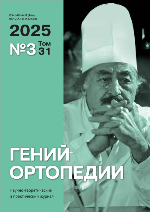
A scientific and practical peer-reviewed medical journal Orthopaedic Genius (Genij Ortopedii) was founded in memory of academician G.A. Ilizarov, an outstanding scientist, professor, pioneering orthopaedic surgeon, honorary member of many foreign academies.

The journal publishes scientific articles, literature reviews, case reports, pilot studies, and new technologies in scientific specialty "traumatology and orthopedics".

The results of fundamental and applied research are based on the principles of evidence-based medicine.

The editorial board welcomes the integrated scientific research, merging of research streams, and interaction of different scientific schools and directions.

The journal provides direct open access to its content, based on the principle of making research results available for the global exchange of knowledge and experience.

The journal is recommended by the Supreme Attestation Board of Russia and is included in the list of leading peer-reviewed scientific journals that publish scientific results of the theses for scientific degrees of doctor and candidate of medical sciences.

Current issue

Vol 31, No 3 (2025)
View or download the full issue PDF (Russian) | PDF

Original articles

269-278 134
Abstract

Introduction Cubitus varus denotes the inward deviation of the supinated forearm on the extended elbow. In cubitus valgus, the forearm is angled away from the body with the arm fully extended. Both deformities manifest clinically as an abnormal carrying angle, along with a cosmetically unsightly appearance, with or without restricted range of motion (ROM).
The aim of the study is to evaluate the results of one-stage supracondylar corrective osteotomy and bone fixation using the Ilizarov apparatus in varus and valgus deformities of the elbow joint, using the angulation‑translation principle (osteotomy rule 2); to determine the effect of this method on the humerus-elbow-wrist angle (HEW), ODD and lateral prominence index (LPI).
Materials and Methods A total of 12 patients, age ranging from 7–24 years, who presented with cubitus varus of ≥ 10° (n = 9) and cubitus valgus (n = 3) of ≥ 20°, were included in the study. All patients underwent acute correction using a mini-incision, supracondylar osteotomy and fixation with the Ilizarov frame.
Results The mean time to union was 14.2 weeks (range, 11–18 weeks). The average duration of follow-up was 24 months. Functional outcome was graded as excellent in 9 cases (75 %), good in 2 (17 %) cases and poor in 1 case (8 %) using the grading system of Oppenheim. For cubitus varus, the mean HEW angle improved significantly, from (–15.5 ± 4.2) pre-operatively to (8.2 ± 1.5) post-operatively. For cubitus valgus, the mean HEW angle was (28.3 ± 5.3) pre-operatively, which improved to (14.1 ± 3.1) post-operatively, which was statistically significant. Complications encountered included superficial pin-tract infection in 1 case, lateral condylar prominence in 1 case and complete radial nerve palsy in 1 case.
Discussion Conventional methods of treatment of cubitus varus or valgus include various corrective osteotomies, typically stabilized with internal fixation. Despite being successful, a substantial number of distressing complications have been reported with the use of internal fixation. The mandatory requirement of post-operative immobilization, resulting in stiffness and disuse atrophy, is a deterrent to the use of internal hardware, which can be easily circumvented by the versatility of the Ilizarov apparatus.
Conclusion External fixation with the Ilizarov apparatus is a versatile means of correction of cubitus varus and valgus. It precisely achieves the desired carrying angle and cosmetic appearance of the elbow. It facilitates residual adjustments in under/over-corrected scenarios. The stability is indisputable. Early joint mobilization leads to an improved functional outcome.

279-286 105
Abstract

Introduction Ankle arthrodesis is a surgical procedure for end-stage arthritis and complex ankle pathologies, offering a limb-salvaging alternative to amputation. This study aims to present clinical experience with the Ilizarov apparatus in achieving stable, painless ankle fusion in patients with varied complex ankle pathologies.
Materials and Methods A retrospective study was conducted involving 27 patients who underwent ankle arthrodesis using the Ilizarov fixator between 2014 and 2024. Clinical and radiological evaluations were performed using the ASAMI scoring system. Surgical techniques, patient demographics, and outcomes were analyzed.
Results All 27 patients achieved successful bone union. ASAMI Bone results were rated excellent in 23 and good in 4. Functional outcomes were rated as good in 22 patients and fair in 5. Pin tract infections were effectively managed with antibiotics. The Ilizarov technique demonstrated superior results in achieving stable, pain-free ankles, even in cases with severe osteomyelitis and destroyed ankles with deformities.
Discussion The Ilizarov apparatus provides a minimally invasive, versatile approach for complex ankle pathologies, enabling dynamic axial compression, early weight-bearing, and deformity correction. Despite limitations such as high costs and skill requirements, its success rate surpasses that of internal fixation techniques.
Conclusion The Ilizarov apparatus is the gold standard for ankle arthrodesis, offering stable fusion and addressing comorbidities such as osteomyelitis and limb length discrepancy, with high patient satisfaction and functional recovery.

287-296 93
Abstract

Introduction The technique of multiapical deformity correction with orthopaedic hexapods can be accepted as "standard." This requires several software calculations a and lengths changes in the 12–18 struts. The "spring" technique was designed to address these disadvantages.
Purpose To analyze the treatment results of patients with lower leg multiapical deformities corrected by "standard" and "spring" techniques
Methods The data of patient group 1 (standard technique, n = 17) and patient group 2 (spring technique, n = 17), were used. Correction accuracy, duration of correction, and fixation periods were compared. In  patients requiring lengthening, fixation and osteosynthesis indices were additionally analyzed. Quality of life and segment function were assessed using the LEFS questionnaire.
Results There was no statistically significant difference between the groups when comparing each of the parameters studied. When comparing LEFS parameters before external fixation (EF) and 2–3 months after  frame removal, a statistically significant difference was observed in patients within each of the first and second groups.
Discussion The correction accuracy in each group was 94.4 %, which, along with the absence of a significant difference in the duration of treatment and data from the LEFS questionnaire, indicates equal clinical effectiveness of both methods. It was noted that treatment with various modifications of circular external fixators is equally uncomfortable for patients. At the same time, the need for only one calculation in a hexapod software instead of 2–3, the ability to change the length of 6 struts instead of 12–18, as well as simpler assembly and less cumbersome construct if there is small (less than 10–12 cm) distance between the rings are advantages of a "spring" technique.
Conclusion Based on the analysis of the criteria used for evaluation, it can be concluded that the “spring” technique for correcting lower leg multiapical deformities is as effective as the “standard” technique. The  advantages of the "spring" technique are associated with the greater convenience of its use: both for the orthopedic surgeon and the patient.

297-306 66
Abstract

Introduction Sagittal morphotypes graded by Roussouly are characterized by specific biomechanics of the spinopelvic alignment (SPA) that can be investigated using the finite element (FE) modeling.     
The objective was to design three-dimensional realistic models simulating anatomical and constitutional types LPA and evaluate deformity and strength of the models under compression.
Materal and methods Lateral standing spondylograms of the skull, pelvis and upper third of the femur were produced for volunteers (n = 169) who agreed to participate in the study. Radiographs were interpreted with  Surgimap 2.3.2.1.) and computed tomography (CT) of the SPA was performed for individuals (n = 5) with average sagittal parameters for each of the five Roussouly morphotypes (I, II, III, IIIA, IV). The CT findings were used to simulate (SolidWorks) five parametric finite element models of normal morphotypes of SPA and examine the deformity and strength.
Results The highest von Mises stresses under compression were measured in the bodies and intervertebral discs (IVD) ThX–LI (2.961 MPa), posterior supporting structures LIV–SI (2.515 Mpa) with type I model; vertebral bodies and IVD of the thoracic and lumbar spine, mainly at the ThXII–LI (3.082 MPa) and LIV– LV (3.120 Mpa) levels with type II model; anterior aspects of the bodies and IVD ThXI–LII, posterior thirds of the bodies, pedicles and facet joints LI–SI (1.720 Mpa) with type III model; the bodies and intervertebral discs of the ThIX–LII vertebrae (1.811 MPa), posterior supporting structures of the LI–SI vertebrae (1.650 Mpa) with type IIIA model; in the spinous processes and articular portion of the arches of the LI–SI vertebrae (3.232 MPa) with type IV model.
Discussion The lateral configuration of the SPA has a key effect on the segmental distribution of gravitational force and determines the specificity of the sagittal biomechanics of the spine, its resistance to dynamic loads and tendency to various degenerative pathologies.
Conclusion Types III and IIIA were the most biomechanically balanced types, hypolordotic form (types  I and II) was associated with overloaded anterior vertebral structures including intervertebral disc protrusion (IDP) and overloaded posterior supporting structures in case of hyperlordosis (type IV).

307-313 69
Abstract

Introduction The concept of the pathogenesis of periprosthetic joint infection (PJI) is the ability of pathogenic microorganisms to colonize the surfaces of implants, which are infected during the surgery or by hematogenous dissemination of bacteria. It causes poor results of PJI treatment. Microbiological identification of pathogen species is the gold standard in the diagnosis of PJI.
Purpose To assess the etiology of the infectious process in patients with periprosthetic hip joint infection.
Methods The study analyzed revision interventions (n = 294) for PJI of the hip joint performed within the period from 2010 throughout 2021. A total of 147 patients were operated on: 56 % (n = 82) were men and  4 % (n = 65) were women. At the time of hospitalization, the fistula PJI type was diagnosed in 71 % (n = 105); 20 % (n = 29) had edema and hyperemia of the postoperative suture area, and 9 % (n = 13) of cases had open wounds.
The object of the study was bone and soft tissue samples obtained during excision of the infected material, as well as removed implant components. Cultures were grown on dense nutrient media. Bacterial cultures were identified by generally accepted methods using TB Expression (BioMerieux, France) and Walk Away 40 (USA) bacteriological analyzers.
Results The etiology of periprosthetic infection was identified in the majority of patients (93 %), while pathogens could not be detected in the remaining cases. Bacteriological analysis revealed microbial associations in 31 % of patients, gram-positive microflora in 52 % of patients, and gram-negative microflora in 10 %.
Discussion The most common types of microorganisms are gram-positive bacteria with a tendency for  resistant strains to grow. Gram-negative bacteria are isolated in joint infection, but less frequently.
The results demonstrate isolated gram-negative cultures in 10 % of cases. The second most common cause of periprosthetic joint infection is polymicrobial infection, which was detected in 31 % of cases. Microbial associations occurr in 10–45 % of cases; such a clinical situation at the start of treatment complicates the empirical choice of drugs for antibacterial therapy.
Conclusions Microbiological study allowed identification of the etiology of the infectious process in 93 % of patients. In more than half of the cases (52 %), the cause of implant-associated infection is grampositive microflora, and in 31 % of cases are microbial associations. Reinfection was noted in 41 % of cases in polymicrobial patients.

314-321 68
Abstract

Introduction Congenital scoliosis is a multifactorial disease caused by abnormalities in vertebral development during embryogenesis. The TBX6 gene, located at locus 16p11.2, plays a key role in  somitogenesis, and  the  heterozygous deletion is associated with the development of specific phenotypes of congenital scoliosis (TBX6-associated congenital scoliosis, TACS). Despite numerous studies on the role of  TBX6 in  the  pathogenesis of congenital scoliosis, there is a paucity of data on the phenotypic manifestations
of heterozygous 16p11.2 deletion.
The objective was to identify and confirm the TACS phenotype being associated with 16p11.2 deletions in the Russian patients.
Material and methods A single-center retrospective cohort study included 187 patients diagnosed with congenital scoliosis treated at the Turner National Medical Research Center for Pediatric Orthopedics and Traumatology between 2012 and 2021. Heterozygous deletion (16p11.2 region) were verified using MQRT‑PCR. The deletion group consisted of 42 patients, and the control group included 145 probands. Clinical and radiological findings were reviewed to identify localization, type and multiplicity of vertebral anomalies and associated malformations. Descriptive statistics and Pearson's correlation coefficient were used for data processing.
Results Heterozygous deletion of TBX6 was detected in 22.4 % of patients. The thoracic and lumbar spine were common localizations, while involvement of the cervical spine was not identified in the deletion group. Vertebral malformations were the most common anomaly in both study groups, but their prevalence was higher among patients with TBX6 deletion (50 % vs. 43.4 %). Multiple spinal malformations were more common in the deletion group (50 % vs. 35 %). Associated internal organ defects were less common in patients with deletion (31 % vs. 43.4 %), while rib synostoses and Sprengel's disease were more common.
Discussion TACS is characterized by specific manifestations including multiple vertebral malformations in the thoracic and lumbar spine, rib synostoses and Sprengel's disease, which is consistent with the scientific literature.
Conclusion The findings indicate the need to include genetic testing for TBX6 deletion in the diagnostic algorithm for congenital scoliosis to facilitate early detection and a personalized approach to treatment of this cohort of patients.

322-333 77
Abstract

Introduction Implant-associated infection (IAI) caused by gram-negative pathogens is characterized by a more severe, recurrent course and higher mortality than the one caused by gram-positive ones. The main reason is growing antibiotic resistance of these pathogens and the complexity of choosing drugs for inpatient and outpatient therapy.
Purpose To evaluate the influence of various factors and compare the features of the course of implant-associated infection caused by P. aeruginosa, K. pneumoniae, A. baumannii in patients with positive and poor treatment outcomes
Methods A retrospective analysis of the medical records of 172 patients treated at the Department of Purulent Osteology between January 1, 2017 and December 31, 2022 for implant-associated infection caused by  P. aeruginosa, K. pneumoniae, A. baumannii was conducted. Based on the results of a telephone survey or examination, patients were divided into 2 groups: positive and poor treatment outcomes by Delphi criteria. The impact of various factors in the anamnesis, laboratory and microbiological analysis, features of surgical intervention, antibacterial therapy and the course of the early postoperative period on the outcomes was analyzed in the IBM SPSS STATISTICS (version 26).
Results Among patients with IAI caused by gram-negative bacteria, the rate of poor outcomes was 45 %, with fatality rate of 10 %. During the comparative study, a statistically significant effect on the development of  a  poor outcome was shown by the postoperative level of serum albumin (p = 0.002), the sensitivity of the isolated isolate to the tested antibacterial drugs (p = 0.011), the isolation of the pathogen from patients’ biomaterial in the postoperative period (p = 0.001), a more frequent need for intravenous administration of albumin and iron (p = 0.003 and p = 0.056, respectively) and the need for repeated surgical intervention in the early postoperative period (p = 0.001).
Discussion IAI caused by gram-negative bacteria is characterized by a prolonged recurrent course and high mortality, primarily associated with the overall growing antibiotic resistance of pathogens which requires an individual approach to both surgical treatment and drug therapy, as well as the development of new tactical approaches to therapy.
Conclusion The rate of poor outcomes was 45 %. Hypoalbuminemia and antibacterial resistance of isolates of P. aeruginosa, K. pneumoniae, A. baumannii, detection of the pathogens in the postoperative material, as well as the need for surgical reoperation in the early postoperative period, are risk factors for poor outcomes.

334-340 57
Abstract

Introduction The use of lysozyme as a bactericidal agent against the leading pathogens of chronic osteomyelitis can become an alternative or supplement to existing antibacterial drugs.

Purpose To study the antibacterial effect of lysozyme against clinical strains of Staphylococcus aureus and Staphylococcus epidermidis

Materials and methods Control strains of Staphylococcus aureus (ATCC 25923), Staphylococcus epidermidis (ATCC 14990) and clinical strains (n = 48), including MRSA (n = 6) and MRSE (n = 6), isolated from wounds and fistulas of patients with chronic osteomyelitis were used as test cultures. The antibacterial effect of lysozyme was assessed using the disk diffusion method.

Results Lysozyme exhibited bactericidal activity against control strains of S. aureus and S. epidermidis, the growth inhibition zone of bacteria was 11–12 mm. Among clinical strains of S. aureus, 87.5 % were sensitive to lysozyme, the growth inhibition zone diameter was 9–13 mm. No bactericidal effect was observed against three strains of S. aureus, including two MRSAs, and continuous bacterial growth was observed around the disk. Among strains of S. epidermidis, the antibacterial activity of lysozyme was observed against 79.2 % of isolates, the growth inhibition diameter was 8–11 mm. Resistance of three MRSE strains to lysozyme was noted. Lysozyme enhanced the effect of vancomycin and cefoxitin against methicillin-sensitive staphylococci and norfloxacin and vancomycin against methicillin-resistant staphylococci.

Discussion Despite the inhibitory effect found, the use of lysozyme alone may be limited due to its possible degradation by proteases, as well as some immunogenicity. There are studies on the synergism of the combined action of lysozyme with various antibiotics on gram-positive and gram-negative bacteria. The data obtained in our experiment showed an increased antibacterial effect by the combined action of antibiotics and lysozyme against the leading causative agents of osteomyelitis.

Conclusion It has been established that lysozyme has an antibacterial effect against clinical strains of S. aureus, S. epidermidis, including MRSA and MRSE, isolated from wounds of patients with chronic osteomyelitis. An increased antibacterial effect is observed by a combined action of lysozyme with cefotaxime, norfloxacin and vancomycin.

341-349 58
Abstract

Introduction Exo-prosthetics of limbs through osseointegration opens up new possibilities in prosthetics. Modern prostheses are becoming more high-tech, which requires deep understanding of the anatomical and functional features of the bone-joint system.
Aim To identify features of structural reorganization of articular cartilage and subchondral zone of the tibia in lower leg prosthetics using an implant with calcium phosphate coating and an implant without additional coating.
Materials and methods The study was performed on 5 intact (control) and 6 experimental dogs (age 1.8 ± 0.5 years, weight 19 ± 1.2 kg). A tibial stump was modeled in the animals at the border of the middle and  upper third of the diaphysis. After 2.5 months a PressFit type implant was installed. Depending on  the  Press‑Fit type, the animals were divided into groups: group 1 made of Ti6Al4V alloy (n = 3); group 2 of Ti6Al4V alloy with calcium phosphate coating (n = 3). Duration of the experiment was 180 days after prosthesis fitting. Histomorphometric study of the articular cartilage and subchondral zone was performed on paraffin sections using an AxioScope.A1 microscope supplied with AxioCam camera and Zenblue software (CarlZeissMicroImagingGmbH, Germany).
Results Bone tissue remodeling was expressed by thinning of the subchondral bone plate, osteolysis, changes in the architecture of bone trabeculae in the subchondral trabecular bone, and a decrease in bone tissue mineralization. These signs were more intense in group 1. Signs of reparative osteogenesis with osteoblasts on the surface of bone trabeculae were noted in group 2. Subchondral bone plate thickness reduced twofold in  group  1, and by 1.5 times in group 2 relative to the control. The values of the parameter of trabecular area were reduced in group 1 by 17 % and in group 2 by 10 %. Statistically significant decrease in the values of  articular cartilage thickness was recorded in group 1 and was accompanied by a higher (by 1.8 times) frequency of vessels been found in the deep zone of cartilage compared to group 2.
Discussion The identified changes in the subchondral zone corresponded to stage 0 (according to the O‑M classification. Aho et al., 2017): very early signs of osteoarthritis, when subchondral sclerosis is not pronounced, the subchondral bone plate is thin. Structural changes in articular cartilage corresponded to  grade  0–1 according to the histological classification of the International Society for the Study of Osteoarthritis OARSI.
Conclusion Histomorphometric changes in the osteochondral component of the tibial plateau during lower leg prosthetics (thinning of the subchondral bone plate, rarefaction of the subchondral trabecular bone, penetration of vessels into non-calcified cartilage) are predictors of arthrosis. The use of implants made of Ti6Al4V alloy coated with a calcium phosphate provides reduction of bone resoption intensity and activates reparative osteogenesis.

350-360 62
Abstract

Introduction The development of new ceramic materials with high osteointegrative characteristics and  experimental substantiation of their application is an important issue in traumatology. The purpose of the work was to study the biological compatibility and osteointegrative characteristics of implants made of zirconium ceramics stabilized with yttrium, ytterbium and gadolinium for filling diaphyseal bone defects in an experiment.
Material and methods The study was performed on 18 male Chinchilla rabbits. Diaphyseal defects with intramedullary implantation of a rod made of a new ceramic porous (PC), non-porous (NPC) material and titanium alloy (TA) were modelled. The animals were divided into 3 groups based on the rod used: PC, NPC and TA (n = 6 in each). Hematological parameters were studied one day before and 8 weeks after the operation.    Withdrawal of animals from the experiment, X-ray control and tissue sampling with subsequent histological and morphometric examination were performed at 8 weeks after the operation. Statistical data processing was performed using the Statistica 10 software. The Kruskal – Wallis test with subsequent intergroup analysis was used to compare the study groups. The Wilcoxon criterion was used to assess changes in dynamics in individual groups. The results are presented as median and interquartile range.
Results Eight weeks after the surgery, in the PC group compared to the NPC and TA groups the levels of  leukocytes, monocytes and granulocytes were significantly lower (p = 0.025; p = 0.022; p = 0.005, respectively); no significant differences were found in other hematological parameters. The results of histomorphological studies showed that better integration of implants was observed when using PC rods compared to TA and  NPC  implants. The thickness of the bone trabecula in the implantation area was
significantly higher in the PC group compared to the TA and NPC groups (86.2 [55.8; 109.9], 56.0 [47.2; 75.9] and 33.1 [19.0; 84.5], respectively, in both cases p < 0.001).
Discussion We studied the biocompatibility and osteointegrative properties of implants made of a new ceramic material in two versions, nonporous and porous (pore size of 10–50 μm), and compared them with titanium alloy implants. It was previously proven that alloyed ceramic materials are attractive for tissue regeneration due to their functional properties, biological activity, and therapeutic effects provided by the introduced ions. The results of our histological and morphometric studies confirmed the better biocompatibility and  osteointegration of implants made of porous zirconium ceramics (PC) containing yttrium, ytterbium, and gadolinium ions, compared to implants made of NPC and TA.
Conclusion A new zirconium-based ceramic demonstrates biological compatibility. Implants with pore sizes of 10–50 μm have good osteointegrative characteristics which determine their possible use in the treatment of bone defects.

361-371 60
Abstract

Introduction Local antibiotic therapy is used to prevent and treat periprosthetic joint infection, but the available antibiotic delivery systems have some limitations.
The objective was to determine optimal parameters of pressure, exposure time and type of solvent to ensure prolonged elution of vancomycin from the original osteosubstituting material based on cancellous allograft bone using an in vitro experiment.
Material and methods Seven impregnation techniques with different combinations of parameters were examined including pressure: from atmospheric to reduced (7–10 hPa), time: from 5 minutes to  24 hours, solvent (distilled water, 50 % ethanol solution, a combination of 50 % ethanol and 5 % polyvinylpyrrolidone (PVP)). The efficacy was assessed by changes in the diameter of the S. aureus ATCC 43300 inhibition zone using  the  bacteriological method and the dynamics of vancomycin concentration
in the eluate and high‑performance liquid chromatography (HPLC). Statistical analysis was performed using the ANOVA method, Tukey's post-hoc test, Spearman's rank correlation and calculation of the area under the pharmacokinetic curve.
Results The best efficiency was demonstrated by the method employing reduced pressure, 60-minute exposure and  an  alcohol solution with PVP, which provided prolonged release of  vancomycin for  14 days with the maximum area under the elution curve (301364.70) and a high correlation between the concentration of  the  antibiotic and the growth inhibition zone (r = 0.908, p < 0.001). The pressure was found to  be the  most  significant factor (F = 19.9916, p < 0.0001), followed by solvent type (F = 7.7485, p = 0.0006) and impregnation time (F = 6.8084, p = 0.0014).
Discussion The technique with use of reduced pressure and an alcohol solution with PVP provides prolonged release of vancomycin for 14 days as opposed to conventional local antibiotic therapy with limited effectiveness of  3 to  7  days. The advantage of the approach includes uniform elution kinetics compared to  polymethyl methacrylate and biodegradable carriers, which demonstrate a sharp initial release of the antibiotic. The complementary use of the microbiological method and HPLC indicated antimicrobial
activity of vancomycin maintained after impregnation being essential for the therapeutic effect.
Conclusion It has been experimentally established that reduced pressure (7–10 hPa), an exposure time of 60 min and the use of 50 % ethanol with 5 % PVP as a solvent appeared to be the optimal parameters for  ensuring prolonged elution of vancomycin from an osteosubstituting material based on cancellous allograft bone.

372-379 61
Abstract

Introduction The established treatments for purulent infection in the bone and joint involve one- or two‑stage local effect on the biofilm with use of bone cement and an active substance including an antibiotic in addition to systemic therapy.
The objective was to evaluate experimental qualitative and quantitative antibiotic release from bone cement introduced into a new type of lattice-structured spacer.
Material and methods A new type of lattice-structured implant/spacer manufactured using additive technologies and a comparison sample simulating a traditional reinforced spacer made of bone cement + antibiotic were used. Vancomycin release was measured by spectrophotometry for periods of 30 days. A regression line was used to plot calibration curves based on data obtained from mother solutions.
Results An effective profile of antibiotic release from bone cement was obtained in the first days of the experiment, followed by a decrease at the end of the first week and an exit to a uniform plateau. The amount of fixed antibiotic in solutions did not exceed 1 % of the total mass of bone cement and active substance. The amount of antibiotic released from the lattice-structured samples was higher than that in the comparison samples.
Discussion Antibiotic release is a superficial process and is not dependent on the total volume of bone cement. A  possible increase in the volume of the medicinal composition does not lead to a proportional increase in the amount of the active substance released. The findings showed that the antibiotic release is more intense even with a smaller volume of material in the lattice structures compared to the control samples, which emphasizes the importance of optimizing the geometry and structure of the material to achieve maximum efficiency of the release of active substances.
Conclusion The lattice structure of implants quantitatively affects the release of antibiotic from bone cement into the environment.

Clinical Case

380-387 55
Abstract

Introduction Atrophic nonunion and defects is a rare complication of clavicle fractures. Therefore questions arise when choosing the optimal method of their treatment.
Purpose We aimed to retrospectively assess the effectiveness of treating atrophic clavicle midshaft nonunion and defects with a free fibular autologous graft fixed with the Ilizarov mini-fixator in combination with an intrameduallary wire.
Materials and methods A retrospective study of 14 patients (11 females, 3 males) in the mean age of 34.1 ± 2.8 years with atrophic nonunion and defects of the clavicle was carried out. Eleven patients had post-traumatic nonunion after failures of its surgical treatment including seven cases of multiple surgeries, and three cases were congenital nonunion. Pain in the clavicle area was the main complaint in 13 patients. Five had minor restrictions in the shoulder joint function, and two had a pronounced adduction contracture of  the  shoulder joint. Surgical treatment included debridement, resection of the ends of  the  fragments to  the  paprika sign, defect plasty with a free autologous fibular graft followed by combined fixation with an intramedullary wire and the Ilizarov mini-fixator. Supportive compression of 1 mm every two weeks was produced at the junction of the fragments in order to stimulate repair. The mini-fixator was removed after radiographic confirmation of a continuous union of the graft with the fragments.
Results and discussion The post-resection defect averaged 3.1 ± 0.2 cm. Union was achieved in 11 cases. The  average period in the mini-fixator was 159.9 ± 11.9 days. In all cases, after dismantling the device, the  range of motion in the shoulder joint retained preoperative parameters. The complications were one graft migration, soft-tissue inflammation and deep infection (two cases). Soft-tissue inflammation was treated with antibiotics while deep infection required prompt debridement. Long-term results were followed in 13 patients. There were no problems with the donor site in the long term. The Ilizarov mini-fixator assisted by an intramedullary wire provides stable fixation and allows compression at the junction of bone fragments with a fibular autograft to stimulate bone formation and union in clavicle midshaft nonunion and defects.
Conclusion The combination of three technical components (autologous grafting, Ilizarov mini-fixator, intramedullary wire) yields positive results in the management of large post-resection defects of the clavicle midshaft. Upon graft consolidation, the clavicle acquires a near-to-normal radiographic bone structure.

Review Article

388-398 86
Abstract

Introduction Smart orthopedic implants integrate advanced sensor technologies to revolutionize joint replacement and orthopedic care. These implants enable real-time monitoring of key parameters such as wear, load distribution, and infection indicators, facilitating early intervention and personalized treatment.
This review aims to evaluate the current advancements, clinical applications, challenges, and future directions of smart orthopedic implants.
Methods A systematic literature review was conducted following PRISMA guidelines, analyzing peer-reviewed studies published between February 2015 and January 2025. Sources were retrieved from PubMed, Scopus, Web of Science, and Google Scholar. Inclusion criteria focused on technological innovations, clinical applications, and regulatory considerations.
Results & Discussion Technological advancements in materials, sensor integration, wireless communication, and artificial intelligence have optimized implant functionality. Smart implants enhance postoperative monitoring, predict implant wear, and personalize rehabilitation. Despite their benefits, challenges such as biocompatibility, data security, battery life, and regulatory approval hinder widespread adoption. Addressing these issues through interdisciplinary research is critical for future developments.
Conclusion Smart orthopedic implants have the potential to transform musculoskeletal healthcare by  enabling real-time patient monitoring and personalized treatment strategies. Continued innovation in materials, AI‑driven analytics, and regulatory frameworks will be crucial for overcoming current limitations and ensuring their widespread clinical adoption.

Anniversary

Necrologue

Announcements

2025-06-10

Список статей, утвержденных для публикации в следующем номере журнала (2025. Т. 31, № 4)

Экспериментальные и теоретические исследования

  1. Экспериментальное топографо-анатомическое обоснование гибридного остеосинтеза малоберцовой кости у пострадавших с переломами лодыжек

Цапенко В.О., Кашанский Ю.Б., Вашетко Р.В., Кондратьев И.П., Поликарпов А.В.

  1. Антистафилококковая активность титановых 3D-имплантатов с магнийсодержащим многокомпонентным покрытием

Гордина Е.М., Божкова С.А., Лабутин Д.В., Богма М.В., Ерузин А.А.

Клинические исследования

  1. Роль микробиологических методов исследования диагностики перипротезной инфекции у больных с асептической нестабильностью эндопротезов тазобедренного сустава

Линник С.А., Оришак Е.А., Ермаков А.М., Кучеев И.О., Нилова Л.Ю., Фадеев Е.М., Карагезов Г., Коршунов Д.Ю., Цололо Я.Б., Усиков В.В., Поликарпов А.В

  1. Clinical and functional outcomes of acute distal tibia fractures treated with ilizarov external fixation: a retrospective study

Manish Dhawan, Brajesh Nandan, Mohammed Schezan Iqbal, Sanjeev Kumar Singh, Manish Prasad

  1. Ошибки и осложнения при устранении посттравматических фронтальных деформаций локтевого сустава методом корригирующей надмыщелковой остеотомии с управлением аппаратом Илизарова

Солдатов Ю.П., Дьячков А.Н.

  1. Результаты хирургической аллопластики дефектов костной ткани различной локализации при переломах дистального отдела плечевой кости

Давыдов А.П., Чибриков А.Г., Ульянов В.Ю., Норкин И.А.

  1. Cравнительный анализ различных модификаций остеосинтеза эластичными стержнями при лечении детей с внесуставными переломами проксимального отдела плечевой кости

Шабанов Д.И., Коробейников А.А.

  1. Валидация видеоассистированного на основе компьютерного зрения гониометрического исследования двигательной функции отведения плечевого сустава

Демкин С.А., Малякина А.А., Ахрамович С.А., Каплунов О.А., Обраменко И.Е., Симонова И.Э.

  1. Гистологические проявления остеоартроза запястья в зависимости от давности и тяжести заболевания

Щудло Н.А., Ступина Т.А., Куттыгул Ш.К.

  1. The incidence and risk factors related to post operative dysphagia after anterior cervical spine surgery: a prospective study

Jagdeep Singh, Navpreet Singh, Pranav Gupta, Anmol Chandhar

Пилотные исследования

  1. Результаты применения внутрисуставных и комбинированных вмешательств у пациентов с ишемической деформацией головки бедра

Тепленький М.П., Бунов В.С., Акимова А.В., Парфёнов Э.М.

Новые технологии

  1. Вариант восстановления плечевой кости при псевдоартрозе свободными аутотрансплантатами малоберцовой кости в условиях чрескостного остеосинтеза

Давиров Ш.М., Борзунов Д.Ю.

Систематические обзоры

  1. Эффективность использования антибактериальных покрытий титановых имплантов при лечении огнестрельных переломов

Давыдов Д.В., Брижань Л.К., Керимов А.А., Хоминец И.В., Бекшоков К.К., Грицюк А.А., Кукушко Е.А., Беседин В.Д.

  1. Специфика ротационного планирования и интраоперационного размещения бедренного компонента эндопротеза коленного сустава с применением навигационных устройств

Зубавленко Р.А., Марков Д.А., Островский В.В.

Клинические наблюдения

  1. Гиперкоррекция оси нижней конечности как исход одномыщелкового эндопротезирования коленного сустава

Корнилов Н.Н., Чугаев Д.В., Иванов П.П., Магомедов М.Ш., Куляба Т.А., Филь А.С.

2025-04-02

Список статей, утвержденных для публикации в следующем номере журнала (2025. Т. 31, № 3)

Экспериментальные и клинические исследования

  1. Экспериментальное изучение условий импрегнации для получения пролонгированной антимикробной активности оригинального остеопластического материала на основе губчатой аллокости

Антипов А.П., Божкова С.А., Гордина Е.М., Гаджимагомедов М.Ш., Кочиш А.А.

  1. Остеоинтегративные характеристики имплантатов из циркониевой керамики и их биологическая совместимость при восполнении диафизарных дефектов

Волокитина Е.А., Саушкин М.В., Антропова И.П., Кутепов С.М., Бриллиант С.А.

  1. Особенности течения и среднесpочные исходы имплантат-ассоциированной инфекции, вызванной ведущими грамотрицательными возбудителями

Туфанова О.С., Божкова С.А., Гордина Е.М., Артюх В.А.

  1. Анализ микробного пейзажа у пациентов с перипротезной инфекцией тазобедренного сустава

Ермаков А.М., Богданова Н.А., Матвеева Е.Л., Гасанова А.Г.

  1. Антибактериальное действие лизоцима против возбудителей остеомиелита: s. Aureus и s. Epidermidis

Шипицына И.В., Осипова Е.В.

  1. Ilizarov ring fixator for ankle fusion: a gold standard in managing complex ankle pathologies

Manish Dhawan, Brajesh Nandan, R K Guhan, Sahil Dwivedi, Manish Prasad

  1. Results of supracondylar osteotomy and bone fixation with the Ilizarov apparatus in the treatment of cubitus varus and valgus deformities

Rajiv Kaul, Manish Prasad, Neha Akhoon

  1. Конечно-элементное моделирование анатомо-конституциональных типов позвоночно-тазового комплекса (Roussouly) в аспекте изучения их биомеханических особенностей

Шульга А.Е., Ульянов В.Ю., Рожкова Ю.Ю., Шувалов С.Д.

  1. Сравнительный анализ коррекции многовершинных деформаций костей голени при помощи различных методик использования ортопедических гексаподов

Головёнкин Е.С., Соломин Л.Н.

  1. Генотип – фенотипическая ассоциация гетерозиготной делеции гена tbx-6 у пациентов с врожденным сколиозом

Хальчицкий С.Е., Виссарионов С.В., Першина П.А., Буслов К.Г., Новосад Ю.А., Согоян М.В., Асадулаев М.С., Герцык М.В.

Пилотные исследования

  1. Апробация эффективности нового типа спейсеров для локальной антибиотикотерапии

Ахтямов И.Ф., Шафигулин Р.А., Галяутдинова А.Э., Харин Н.В., Беспалов И.А., Бойчук С.В., Саченков О.А.

Клинические наблюдения

  1. Серия клинических наблюдений лечения пациентов с гипотрофическими псевдоартрозами и дефектами диафиза ключицы с применением свободной аутопластики трансплантатом малоберцовой кости, минификсатора Илизарова и интрамедуллярного армирования

Колчин С.Н., Моховиков Д.С., Малкова Т.А.

Систематический обзор

  1. Smart orthopedic implants: the future of personalized joint replacement and monitoring

Kirolos Eskandar

2025-02-06

Список статей, утвержденных для публикации в следующем номере журнала (2025. Т. 31, № 2)

Экспериментальные исследования

  1. Экспериментальная оценка свойств костнозамещающих материалов на основе CA3(PO₄)₂ на модели критического дефекта бедренной кости крысы
    Щербаков И.М., Евдокимов П.В., Ларионов Д.С., Путляев В.И., Шипунов Г.А., Данилова Н.В., Ефименко А.Ю., Новоселецкая Е.С., Дубров В.Э.

Клинические исследования

  1. Факторы риска нестабильности имплантатов после спондилэктомии у пациентов с опухолями позвоночника
    Заборовский Н.С., Масевнин С.В., Мураховский В.С., Мухиддинов В.С., Смекалёнков О.А., Пташников Д.А.
  2. Этиология инфекционного спондилодисцита: есть ли связь с успехом лечения
    Любимова Л.В., Любимов Е.А., Павлова С.И.
  3. The diagnostic value of IL-6 and thymidine phosphorylase in the progression of osteosarcoma
    Abdul Azeez B.A., Abd Alhussain G.S., Salman R.S., Al-Fahham A.A.
  4. Definitive fixation of open tibial fractures using Ilizarov ring fixators-an analysis of functional outcome
    Radhakrishnan E., Duraisamy E.
  5. Лечение детей с политравмой таза методиками малоинвазивного и комбинированного остеосинтеза
    Борозда И.В., Николаев Р.В., Каримов М.Ю., Салахиддинов Ф.Б., Машарипов Ф.А.
  6. Зависимость уровня сывороточного прокальцитонина от микрофлоры в очаге инфекции при хроническом остеомиелите
    Шипицына И.В., Осипова Е.В., Шастов А.Л.
  7. Сравнительный анализ исходов хирургического лечения пациентов после артродеза и подвешивающей артропластики седловидного сустава
    Афанасьев А.О., Чижов А.Е., Абдиба Н.В., Родоманова Л.А.
  8. Способ прогнозирования исхода оперативного лечения контрактуры Дюпюитрена на основе показателей лейкоформулы
    Щудло Н.А., Сбродова Л.И., Останина Д.А.
  9. Клинико-статистический анализ состояния культей как элемент выявления противопоказаний к протезированию
    Пепеляев А.В., Скоблин А.А.
  10. Кинематические и кинетические особенности не вовлеченной конечности при ходьбе у детей со спастической гемиплегией
    Мамедов У.Ф., Долганова Т.И., Смолькова Л.В., Трофимов А.О., Гатамов О.И., Попков Д.А.

Пилотные исследования

  1. Прогнозирование нарушения консолидации переломов длинных костей конечностей с помощью нейросетевого анализа
    Мироманов А.М., Гусев К.А., Старосельников А.Н., Мудров В.А.

Мета-анализ

  1. Proximal femoral nail antirotation versus bipolar hemiarthroplasty for intertrochanteric fractures
    I Gusti Agung Ditya Damara, Nyoman Satria Nakayoshi Wijaya, I Wayan Suryanto Dusak
More Announcements...


Creative Commons License
This work is licensed under a Creative Commons Attribution 4.0 License.